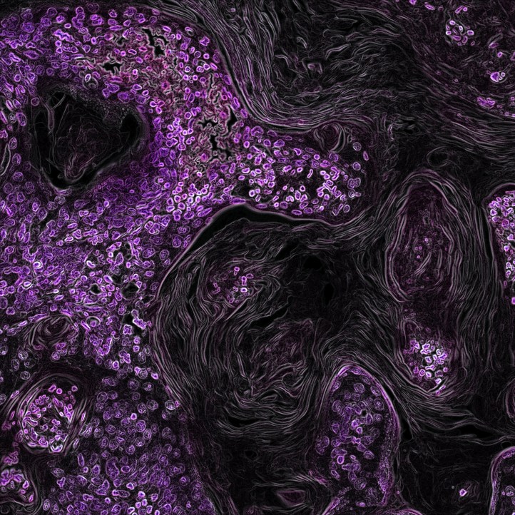
 There’s a strange beauty to these technically superb visualizations of cancer. In the image above, a purple label marks lung cancer cells driven by the Kras oncogene in a genetically engineered mouse.
Here, a breast tumor is imaged by multiphoton microscopy and endogenous fluorescence, i.e., without the use of staining.
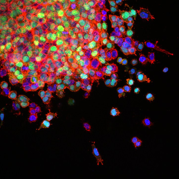
In this view of a metastasizing lung cancer, green fluorescent protein labels a gene mutation that promotes the metastatic cascade. The cytoskeletal protein actin is red and cell nuclei are blue.

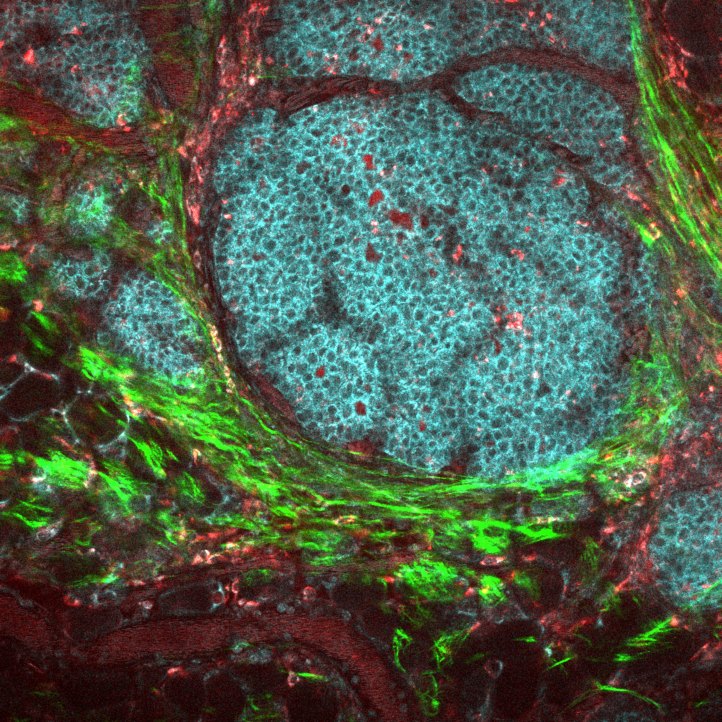
A technique called transparent tumor tomography shows a HER2-positive breast cancer at a single-cell resolution. HER2 is green, Ki-67 is red, PD-L1 is purple, immune cells are yellow, and endothelial cells cyan. This is just a sample from the National Cancer Institute’s Cancer Close Up online exhibit.

Great post thank yoou